


Special tests
Although not a routine test, ultrasound (scan) can convey the following information:
- Confirm that pregnancy is in the uterus and not in the tubes.
- Confirm the number of foetuses.
- Help to estimate gestational age of foetus.
- Check for foetal malformations (at 20 weeks).
- Help establish position and condition of foetus and placenta (in late pregnancy).
Some caregivers perform a scan at every visit; others are more conservative in their approach and may scan less often. The guideline below indicates what a scan will show at various times during your pregnancy.
Early pregnancy scan
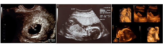
Week 5: The pregnancy sac is visible.
Weeks 6–7: The embryo can be seen and the heart movement is visible.
Week 8: Early foetal movements and twins can be confirmed.
Weeks 9–10: The outline of the placenta is visible and the first measurements of the foetus can be made to establish a due date.
Midway scanning
Weeks 16–26: The foetus is larger and the organs become visible. The baby’s head is measured and femur length determined – both are important for determining the age of the foetus. Other important structures looked for in a routine scan include the stomach, the bladder, the spine and the external genitalia. The placenta is easily recognised from the rest of the uterus. The amount of amniotic fluid can also be assessed.
A scan done between 18–24 weeks is the single most important routine scan, because it gives an accurate assessment of the gestational age and can detect most of the major abnormalities.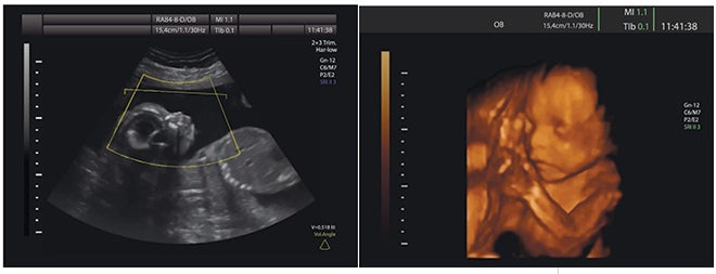
Late pregnancy scanning
The main reason for scanning at this late stage is to monitor foetal growth. This is done by measuring the circumference of the baby’s head, the length of the femur and the size of the abdomen. If there is any concern regarding foetal wellbeing, a cardiotocograph (CTG) will be done together with an assessment of foetal movements.
Related articles

Further comfort measures for pain relief
Distractions like breathing, visualising or fixing on one point can take your mind off the pain.
5 mins to read

Levels of fitness
First trimester
Your fitness is likely to decrease. Listen to your body and only do what you feel comfortable doing.
Second trimester
5 mins to read

Monitoring your response to exercise
It is important to monitor your response to your exercise programme by taking your pulse-rate before, during and after exercise.
5 mins to read

Keys to relaxation
Physical environment
5 mins to read

Criteria for prenatal classes
Instructor’s credentials, for example, midwife, physiotherapist.
Instructor’s specialised training in childbirth education.
5 mins to read

Conception
The fertilised egg divides into two identical cells – then four, then eight, then 16, and then many billions, and 266 days later – your baby.
1 min to read

Exercise
Feeling healthy, stress-free and relaxed will only help the chance of you and your partner falling pregnant.
1 min to read

Increasing postural awareness and correction take
Take particular care of your back during and after pregnancy – protect it from strain and possible injury.
5 mins to read

Adjusting to pregnancy
Discovering that you are pregnant brings about a variety of new feelings.
5 mins to read

Hazards to pregnancy
Pregnancy is usually diagnosed several weeks after conception. It is, therefore, advisable to avoid risk factors if you are planning to become pregnant.
2 mins to read

Maintaining and improving fitness
Stimulating the cardiovascular system through aerobic exercise helps to maintain and improve your fitness during pregnancy.
5 mins to read

Types of relaxation techniques
Progressive relaxation
2 mins to read

How Much Weight Should I Gain During Pregnancy?
There are many things you can do to prepare for the delivery of a healthy baby. One of the most important things is eating right to gain the extra weight you’ll need to support another life.
4 mins to read

Preconception checklist
Are you planning to have a baby soon? Find out our preconception checklist to facilitate the conceiving process and have an healthier pregnancy.
1 min to read

Energy and weight
Energy
You need extra energy:
5 mins to read

Breathing techniques
Breathing reflects the activity in your body and, of all the body’s involuntary unconscious physical responses, breathing is the easiest to control.
4 mins to read

Sex of the Baby
Women have XX and men have XY chromosomes.
1 min to read

Diagnostic tests
Diagnostic ultrasound
The most important scans are performed at 12 weeks and 16 weeks. These early scans often prove the most accurate in determining the date of birth.
3 mins to read
Relaxation and correct breathing techniques
As your pregnancy advances, the uterus exerts an increased upward pressure on your diaphragm. As you near your due date you may find that you breathe less deeply, although more efficiently.
5 mins to read